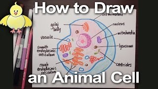
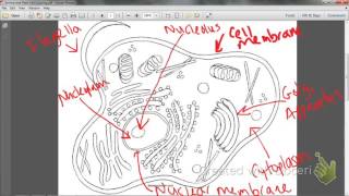
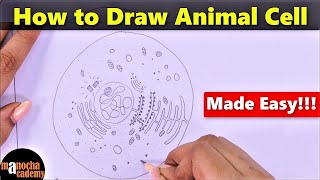
Animal Cell Coloring With Labels
Hi friends, welcome to a fun and educational activity that brings the microscopic world to life. In this guide we’ll walk through how to draw an animal cell, color it beautifully, and add clear labels so you can remember every part. Whether you’re a student studying biology, a teacher preparing a classroom activity, or simply curious about the building blocks of life, this step‑by‑step tutorial will help you create a vivid, accurate representation of an animal cell.
Why Color and Label an Animal Cell?
Coloring a cell is more than a creative pastime. By assigning distinct hues to each organelle, you reinforce memory and gain a deeper appreciation for how these structures work together. Labels clarify function, making the drawing a useful reference for exams, presentations, or science projects.
Materials Needed
- Sketchpad or heavy paper
- Pencils (HB for sketching, 2B for shading)
- Eraser
- Colored pencils, markers, or watercolors
- Fine‑tip pen or ink for outlining
- Reference image of an animal cell (optional)
Step 1: Outline the Cell Boundary
Begin by drawing a large, slightly irregular oval to represent the plasma membrane. Use a light pencil stroke so you can adjust the shape as you add organelles. The membrane is the outer boundary that controls what enters and leaves the cell.
Step 2: Add the Cytoplasm
Inside the oval, shade a gentle, translucent layer to mimic cytoplasm. This jelly‑like substance houses all organelles and provides a medium for chemical reactions. Keep the shading light so that the organelles you add later stand out.
Step 3: Draw the Nucleus
Place a large circle near the center of the cell. Inside the nucleus, add a smaller, darker circle to represent the nucleolus. The nucleus controls cell activities, while the nucleolus is where ribosomal RNA is produced.
Step 4: Place the Endoplasmic Reticulum
Sketch two types of endoplasmic reticulum (ER): rough and smooth. Rough ER consists of ribosomes attached to its surface; draw tiny dots along its sheets. Smooth ER has a smooth appearance; draw it as a series of connected tubes. ER is responsible for protein synthesis (rough) and lipid metabolism (smooth).
Step 5: Add the Golgi Apparatus
Draw a stack of flattened sacs near the ER. The Golgi apparatus modifies, sorts, and packages proteins and lipids for transport. A simple stack of three to five rectangles will convey its structure.
Step 6: Include the Mitochondria
Insert two or three oval shapes with cristae—folded inner membranes—inside the cytoplasm. Mitochondria generate ATP, the cell’s