Curated coverage and comprehensive guides by Fresh Picks.
# Establishing a Systematic Approach to CT Chest Interpretation
Interpretation of CT chest examinations is a fundamental skill for any radiology trainee. For first year residents starting out on their clinical rotations, the volume of information contained in a single chest CT can be overwhelming. Without a structured method, it is easy to miss key findings or misinterpret normal anatomy. Developing a consistent system early in training builds confidence, reduces errors, and ensures that every scan is reviewed comprehensively.
## Why a System Matters in CT Chest Interpretation
A key anatomic feature of the lung that determines how lung pathology appears on CT is the secondary pulmonary lobule. Understanding this basic unit helps differentiate between airspace, interstitial, and airway diseases. But anatomy alone is not enough. When faced with a complex case, having a step‑by‑step routine allows the reader to evaluate each component of the examination without overlooking subtle abnormalities.
Many educators stress that having a system to go through each kind of scan is so important—without it, it is so easy to miss things. This is especially true in chest CT, where findings range from small pulmonary nodules to subtle ground‑glass opacities. A systematic approach standardizes the review process and provides a framework that can be applied to both routine and emergency studies.
## Building a Practical Checklist
A useful system for CT chest interpretation can be organised into several consecutive steps. Begin with patient demographics and the indication for the study. This context directs attention to the most likely pathologies. Next, assess the trachea and central airways, then evaluate the lung parenchyma using lung windows. After that, examine the mediastinum using soft tissue windows, followed by the pleura, chest wall, and upper abdomen. Finally, review any incidental findings.
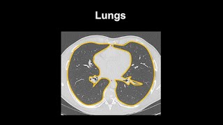
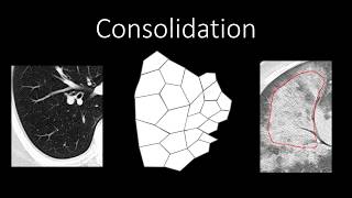
Within the lung parenchyma, compare side to side for symmetry. Look for nodules, consolidations, ground‑glass opacities, reticular patterns, and cysts. The interpreter should describe the distribution—central, peripheral, upper lobe predominant—and characterise each abnormality. Using consistent terminology helps communicate findings clearly to referring clinicians.
## Applying the System to Cases
In an educational video on CT chest interpretation, Dr. Jannette Collins presents multiple cases to demonstrate how a systematic approach works in practice. The session includes a structured timeline: introduction, then sequential cases that build in complexity. For example, Case 1 might focus on a simple solitary pulmonary nodule, while later cases introduce interstitial lung disease, infection, and neoplasm. This progression helps viewers apply their systematic checklist to increasingly challenging scenarios.
Learners who could not attend a live lecture can still benefit from such recorded resources. Following along with cases reinforces the habit of using a consistent method. It also allows the viewer to pause and review specific findings, which is especially helpful for first year residents who are still learning the spectrum of normal variants.
## Common Pitfalls for Beginners
One of the most common errors in early CT chest interpretation is the failure to
Related Coverage
Introduction to CT Chest - Anatomy and Approach
Access our
How I Read a Chest CT
This is for first year residents, starting out on their
How to Read a Chest CT
When radiologists read a
How to Review a Chest CT: My Search Pattern
Having a system to go through each kind of scan is so important - without it, it is so easy to miss things! Here I go through my ...
How to identify normal lung anatomy on chest CT
A key anatomic feature of the lung that determines how lung pathology appears on
CT Patterns of Lung Disease, Dr. Jannette Collins - Medality (MRI Online) Radiology Noon Conference
In this video, Dr. Jannette Collins presents
Chest CT Patterns of Interstitial Lung Disease
In this presentation, Dr. Elsie Nguyen, an associate professor of radiology at the University of Toronto, discusses the patterns of ...
CHEST CT IN ICU PATIENTS: DR R GOTHI
00:00:00 INTRO 00:04:25 CASE1 00:06:22 CASE 2 00:09:54 CASE 3 00:12:15 CASE 4 00:13:04 CASE 5 00:16:26 CASE 6 ...
Chest Radiology - Patterns - Airspace
Audience: Medical Students and Residents Learning Objectives: Differentiate airspace and interstitial opacities Describe the ...
CHEST 2016 video sessions: HRCT of the chest
Could not attend
Classic Signs | Chest Radiology Board Review
Classic radiographic signs on CXR and
Chest X-Ray and Chest CT Interpretation
A case covering
Anatomy of a Transverse CT of the Thorax
An overview of the anatomy visible in a transverse computed axial tomographical image of the